Offer
Provide additional details about the offer you're running.
FLASHSALE

Time to read 12 min
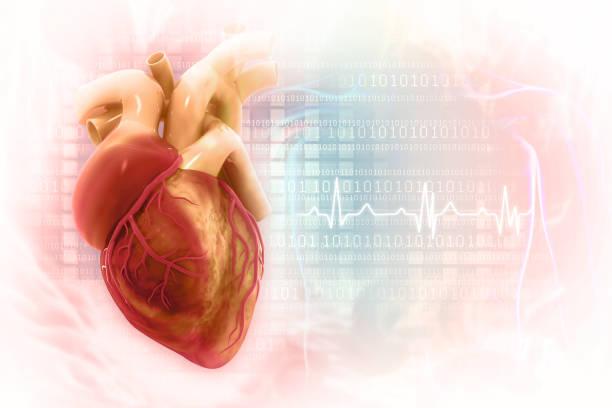
An ECG (Electrocardiogram) test helps detect several common heart problems by recording the heart’s electrical activity. It can identify irregular heartbeats (arrhythmias), signs of a heart attack, poor blood flow to the heart, enlarged heart chambers, and conduction abnormalities. Doctors also use ECGs in different heart diseases to monitor existing heart conditions and evaluate symptoms such as chest pain, dizziness, shortness of breath, or palpitations. Since the test is quick, painless, and non-invasive, it is widely used for early detection of potential heart issues.
An electrocardiogram is a clinical test that determines the heart's electrical activity. The test is not invasive. An ECG can help your doctor diagnose heart disease, a heart attack, or a lack of blood supply to the heart by analyzing its electrical signals. The purpose of this article is to explain how ECG tests help detect heart problems early.
According to the American Heart Association, ECG is one of the most commonly used diagnostic tools for detecting arrhythmias and signs of heart attack. An ECG (EKG) can help detect many heart problems, including Arrhythmia, Coronary Artery Disease, heart attacks, and structural heart abnormalities.
An ECG (Electrocardiogram) test is a simple and painless procedure that records the electrical activity of the heart. It helps doctors evaluate heart rhythm, heart rate, and overall heart function. ECG tests are commonly used to detect conditions such as irregular heartbeat, heart attack, blocked arteries, and other cardiac abnormalities. The test is quick, non-invasive, and often recommended when a person experiences symptoms like chest pain, palpitations, dizziness, or shortness of breath.
|
Problems |
How Electrocardiography Can Help |
|---|---|
|
Atrial fibrillation |
ECG shows an irregular and often rapid heartbeat without clear P waves |
|
Ventricular tachycardia |
ECG detects a dangerously fast rhythm originating from the ventricles |
|
Sinus tachycardia |
ECG shows a fast but regular rhythm from the heart’s natural pacemaker |
|
Bundle branch block |
ECG reveals delayed electrical conduction through the heart’s ventricles |
|
Cardiac ischemia |
ECG may show ST depression or T-wave inversion due to reduced blood flow |
|
Angina |
ECG can show temporary ischemic changes during chest pain episodes |
|
Cardiac arrest |
ECG may display life-threatening rhythms like ventricular fibrillation or asystole |
|
Atrial flutter |
ECG shows rapid, organized atrial activity with characteristic flutter waves |
|
Premature ventricular contractions (PVCs) |
ECG records extra abnormal beats arising from the ventricles |
An ECG analyses electrical signals to help your doctor determine how well your heart functions. It monitors irregular heartbeats. It is a tool in cardiology for detecting heart disease and arrhythmias. It also helps you detect signs of previous or impending heart attacks.
An electrocardiogram (ECG) is a simple and non-invasive test that measures the electrical activity of your heart. Here's how:
Preparation: At the clinic, they will lie you down. Then the electrodes are placed on your chest, arms, and legs. These small patches help you to monitor your heartbeat.
Recording: The electrodes detect electrical signals produced by your heart, which are recorded by the ECG machine.
At MyDiagnostics, we support preventive heart health through advanced diagnostic testing and timely health screening services.
|
Heart Disease / Condition |
Common ECG Findings |
|---|---|
|
Arrhythmia |
Irregular rhythm, abnormal heart rate, premature beats, atrial fibrillation, or tachycardia patterns |
|
Coronary Artery Disease (CAD) |
ST-segment depression, T-wave inversion, and signs of reduced blood supply to the heart |
|
Myocardial Infarction (Heart Attack) |
ST-segment elevation or depression, pathological Q waves, T-wave inversion |
|
Heart Failure |
Sinus tachycardia, atrial fibrillation, ventricular hypertrophy, conduction abnormalities |
|
Pericarditis |
Diffuse ST-segment elevation, PR-segment depression |
|
Left Ventricular Hypertrophy (LVH) |
Increased QRS voltage, left axis deviation, strain pattern with ST-T changes |
|
Conduction Disorders |
Prolonged PR interval, widened QRS complex, bundle branch block, heart block pattern. |
Yes. An ECG can detect abnormal heart rhythms, signs of heart attack, poor blood flow, enlarged heart chambers, and electrical conduction problems. However, some heart conditions may require additional tests for confirmation.
|
Health Problem |
Description |
Common ECG Findings |
|---|---|---|
|
Arrhythmias |
Irregular or abnormal heart rhythm |
Irregular heartbeat patterns, abnormal rate |
|
Ischemic Heart Disease |
Reduced blood flow to the heart muscle |
ST-segment depression or T-wave changes |
|
Heart Attack (Myocardial Infarction) |
Blocked blood supply causing heart muscle damage |
ST elevation, abnormal Q waves |
|
Left Ventricular Hypertrophy (LVH) |
Thickening of the heart’s left pumping chamber |
Increased QRS voltage patterns |
|
Heart Block |
Delay or blockage in electrical signal conduction |
Prolonged PR interval or dropped beats |
|
Pericarditis |
Inflammation of the heart’s outer lining |
Widespread ST elevation, PR depression |
Here are the most common cardiovascular problems that you should be aware of:
This condition causes the heart to beat irregularly. An ECG uses specific waveforms, such as absent P waves or erratic R-R intervals, to determine arrhythmia. Atrial fibrillation, for example, is characterized by the absence of a distinct P wave, whereas ventricular tachycardia is identified by rapid, wide QRS complexes on the ECG.
Ischemic heart disease is a cardiovascular condition caused by blocked arteries. Narrowing of the channels reduces blood flow to the heart muscle. This can result in ischemia, which is when the heart muscle is deprived of oxygen, or even a heart attack.
Suppose there is a blockage in the blood supply to a portion of the heart. It kills the affected heart muscle, resulting in a heart attack. Clinical ECGs reveal specific changes in the heart's electrical activity. ST-segment elevation may indicate acute myocardial injury or heart attack. The T-wave inversion and pathologic Q waves indicate heart damage.
The thickening of the heart’s left ventricle is often due to high blood pressure or other underlying conditions. This makes it harder to pump blood. Thus increasing the risk of heart failure.
This occurs when electrical signals in the heart are delayed or completely blocked from the atria to the ventricles. These are classified into three types:
First-degree: This condition is indicated by an ECG with a prolonged PR interval. First-degree heart block occurs when electrical signals are delayed but still reach the ventricles.
Second-degree: This heart block is defined as intermittent failure of electrical signals to reach the ventricles. This results in missing beats.
Third-degree: In third-degree heart block, no signals reach the ventricles, causing the atria and ventricles to function independently.
This is an inflammation of the pericardium, the sac-like covering of the heart. This condition often causes sharp chest pain that worsens with deep breathing or lying down. An ECG is essential in diagnosing pericarditis, as it reveals distinctive changes in the heart’s electrical activity.
One of the hallmark ECG findings in pericarditis is ST-segment elevation, which differs from the localized elevation seen in a heart attack. In some cases, the test may also show PR-segment depression. This indicates inflammation of the pericardium. Early diagnosis of pericarditis helps reduce the risk of complications such as cardiac tamponade.
The electrocardiogram (ECG) is an important tool for detecting heart problems. An ECG measures the electrical signals, providing invaluable insights into its functionality.
This test supports early detection and timely intervention and significantly improve patient outcomes. For example, heart disease frequently develops silently, and regular ECGs can detect issues such as arrhythmias or signs of ischemia before they progress to more serious complications such as heart attacks or strokes.
|
Disease |
Electrocardiography Finding |
|---|---|
|
Heart Attack |
ST elevation |
|
Heart Block |
Prolonged PR interval |
|
Arrhythmia |
Irregular rhythm |
|
LVH (Left Ventricular Hypertrophy) |
Increased QRS voltage |
Early detection is critical because many heart conditions can cause serious health problems if left untreated. An ECG can detect irregular heartbeats, indicating underlying issues that may necessitate lifestyle changes, medications, or surgical procedures. This proactive approach helps patients manage their heart health more effectively and lowers the risk of sudden cardiac events.
While ECGs are extremely effective, they are frequently used in conjunction with other diagnostic tests to provide a more complete assessment of heart health.
Echocardiograms reveal information about the function of the heart. Stress tests assess the heart's response to physical exertion.
The role of ECGs is also a crucial component of preventive cardiology. By assessing family history, high blood pressure, and diabetes, routine ECG screenings are mandatory for at-risk patients. These screenings help with lifestyle modifications before any symptoms appear.
Yes, an ECG can sometimes appear normal even when a person has heart disease. Some heart problems, especially mild or partial coronary artery blockages, may not cause visible ECG changes at rest. In many cases, symptoms occur only during physical activity or stress.
An ECG mainly records the heart’s electrical activity, so it may not always detect the exact location or severity of blocked arteries. Because of this, additional tests such as stress ECG, echocardiography, CT scan, or coronary angiography may be needed for a more accurate diagnosis.
A normal ECG does not always completely rule out heart disease, especially if symptoms like chest pain, breathlessness, or palpitations continue.
There are several ways to lower your risk of developing coronary heart disease (CHD), including lowering your blood pressure and cholesterol levels.
A low-fat, high-fiber diet is recommended, with plenty of fresh fruits and vegetables (5 portions per day) and whole grains.
You should limit your salt intake to no more than 6 g per day, as excess salt will raise your blood pressure. 6g of salt is approximately 1 teaspoonful.
There are two types of fats: saturated and unsaturated. You should avoid foods high in saturated fat because they raise the levels of bad cholesterol in your blood.
The following foods are high in saturated fat:
Includes meat pies, sausages, and fatty cuts of meat.
The following foods are high in saturated fat:
Include meat pies, sausages, and fatty cuts of meat with butter.
Ghee is a type of butter commonly used in Indian cooking. Other ingredients include lard, cream, hard cheese, cakes and biscuits, and coconut or palm oil.
A balanced diet, however, should include unsaturated fats, which have been shown to raise good cholesterol levels and help reduce artery blockages.
The following foods are high in unsaturated fat:
Oily fish, avocados, nuts, and seeds.
Sunflower, Rapeseed, Olive, and Vegetable oils
You should also try to limit your sugar intake, as this can increase your chances of developing diabetes, which has been linked to an increased risk of CHD.
A healthy diet with regular exercise is the most effective way to maintain a healthy weight. A healthy weight reduces your risk of developing high blood pressure.
Regular exercise helps to improve the efficiency of your heart and blood circulatory system, lowers cholesterol, and maintains healthy blood pressure.
Regular exercise reduces your risk of having a heart attack. The heart is a muscle like any other, and it benefits from exercise. A strong heart can move more blood throughout your body with less effort.
Any aerobic exercise, such as walking, swimming, and dancing, works your heart harder and keeps it healthy.
A general practitioner or practice nurse can advise you on your ideal weight based on your height and build. Alternatively, you can get your body mass index (BM) online.
If you smoke, quitting will lower your risk of developing CHD. Smoking is a significant risk factor for developing atherosclerosis.
According to research, smoking medications such as patches or gum increase your chances of successfully quitting smoking by three times.
If you drink, do not go over the maximum recommended limit. If you drink up to 14 units per week, spread it out over at least three days.
If you want to cut back, try having several drink-free days each week. Always avoid binge drinking, as it raises the risk of a heart attack.
You can control your blood pressure by eating a low-saturated fat diet, exercising regularly, and, if necessary, taking blood pressure medications. Your target blood pressure should be lower than 135/85 mmHg. If you have high blood pressure, ask your doctor to check it regularly.
Diabetes increases your risk of developing coronary heart disease. Physical activity and weight control can help reduce your risk of heart problems. It's also critical to have your blood pressure, cholesterol, and blood sugar levels checked regularly. If you have diabetes and are under the age of 80, your target blood pressure should be less than 140/90 mmHg.
If you have severe issues of the heart, you may be prescribed medication to alleviate your symptoms and prevent further complications. If you do not have chronic conditions of the heart but have high cholesterol, high blood pressure, or a family history of heart disease, your doctor may prescribe medication to help you avoid developing heart problems. If you have been prescribed medication, you must take it and adhere to the proper dosage.
You may need an ECG if you experience symptoms or risk factors that could indicate a heart problem, including:
Chest pain or pressure
Palpitations or irregular heartbeat
Dizziness or fainting episodes
Shortness of breath
Family history of heart disease
Diabetes or high blood sugar
Hypertension (high blood pressure)
Unexplained fatigue or weakness
An ECG helps doctors evaluate the heart’s electrical activity and detect possible rhythm problems, reduced blood flow, or signs of heart disease.
ECG technology helps monitor your overall health, but it's important to lead a healthy lifestyle. It can prevent heart disease. Combining ECG with other tests helps catch heart problems early, and it's important for doctors to keep an eye on patients' conditions. Regular ECG monitoring allows doctors to advise on lifestyle changes and heart health. Without ECG.
Healthcare providers can give personalized care by using ECGs along with other tests and new technology. Keeping track of your heart with regular ECG monitoring helps you make choices to prevent serious heart problems in the future.
If you're looking for cardiac specialists or clinics offering ECG services, consult your local healthcare provider or search for cardiology clinics in your area. They can provide you with contact information and schedule appointments for ECG testing.
Yes, Electrocardiography can show many heart problems, including abnormal heart rhythms, reduced blood flow, heart enlargement, and signs of a heart attack, though additional tests may sometimes be needed for confirmation.
Yes, the test can detect signs of heart blockage in the electrical conduction system. It exhibits abnormal rhythms or patterns.
A combination of regular heart check-ups, including an ECG, physical exams, and lifestyle assessments, can aid in assessing heart health. Monitoring for symptoms such as chest pain or shortness of breath is also necessary.
Common symptoms include chest pain, shortness of breath, fatigue, dizziness, and an irregular heartbeat. If you experience any of the following, seek immediate medical attention.
These diseases can be diagnosed through ECGs, echocardiograms, and blood tests. Sometimes a stress test is also performed with these. A review of medical history and symptoms can also detect cardiovascular diseases.
Arrhythmias or evidence of previous heart attacks are associated with cardiovascular abnormalities. An ECG can detect irregularities indicating heart disease.
While an ECG can indicate the presence of a heart blockage, additional tests such as stress tests or coronary angiography may be required to confirm the diagnosis.
Yes, an ECG can sometimes detect signs of Coronary Artery Disease by showing changes related to reduced blood flow or previous heart damage. However, a normal ECG does not always rule out coronary artery disease, so additional tests may be needed for accurate diagnosis.
*** Medical Disclaimer: The following information is for educational purposes only. No information provided on this website, including text, graphics, and images, is intended as a substitute for professional medical advice. Please consult with your doctor about specific medical advice about your condition(s).


