Offer
Provide additional details about the offer you're running.
FLASHSALE

Time to read 15 min
Table of contents
The main difference between an ECG and an echocardiogram is that an ECG records the heart’s electrical activity, while an echocardiogram uses ultrasound to create images of the heart’s structure and blood flow. ECGs are mainly used to detect rhythm problems, whereas echocardiograms help diagnose valve disease, heart failure, and structural abnormalities.
Cardiac diagnosis is basic in the identification, prevention, and management of cardiovascular disease (CVD), which remains the leading cause of mortality in the world.
Because of the complexity of the heart and its operations, early detection of such problems is crucial in preventing them from developing into a life-threatening form.
This is where more detailed diagnostic tools like the electrocardiogram (ECG) and the echocardiogram prove useful. Such tests help doctors observe the heart's performance, identify complications, and suggest personalized therapy depending on the patient’s condition. Both an ECG and an Echo are non-invasive diagnostic tools that do not emit radiation.
The World Health Organization estimates that cardiovascular diseases are responsible for about 17.9 million fatalities. The majority of these deaths occur through heart attacks and strokes that are the result of undiagnosed or poorly managed cardiac issues.
This is why annual cardiac examinations and other diagnostic procedures, including ECG and echo, should be performed not only for patients with risk factors for cardiovascular diseases like hypertension, diabetes, or a family history of heart disease, but also for any asymptomatic patients who can have latent heart disease. These diagnostic tools play vital roles in assessing heart health and are often used during routine check-ups as part of a patient’s heart health journey.
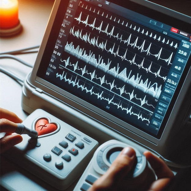
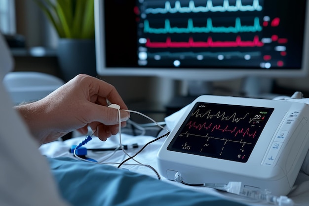
An ECG (electrocardiogram) is an external process in which the electrical signals of the heart are measured over a short span, normally just several seconds. The test offers details on the heart’s rhythm and rate, and any abnormality in the organ’s electrical activity.
It is especially helpful in establishing the diagnosis of arrhythmias, ischemia, and any other issues with the heart’s electrical conduction system.
On the other hand, an echocardiogram, often called “echo,” is a test that employs high-frequency sound waves in the formation of real-time images of the heart. This particular imaging test helps give an idea of the size and location of the heart’s chambers and valves, as well as how blood circulates through the chambers.
It is particularly useful in identifying structural disorders like valve disorders, congestive heart failure, and congenital diseases and assessing general heart function.
In this detailed article, we look into two diagnostic tests, starting with an overview of their methods, then their applications, pros, cons, and the contexts within which they are most relevant, and so on.
The focus is to provide the readers with the idea about when each test is employed, what the tests can reveal, and what the patients should expect during the tests.
On the other hand, an echocardiogram, often called “echo,” is a test that employs sound waves in the formation of real-time images of the heart. This particular imaging test helps give an idea of the size and location of the heart’s chambers and valves, as well as how blood circulates through the chambers. It is particularly useful in identifying structural disorders like valve disorders, congestive heart failure, and congenital diseases and assessing general heart function.
In this detailed article, we look into two diagnostic tests, starting with an overview of their methods, then their applications, pros, cons, and the contexts within which they are most relevant, and so on. The focus is to provide the readers with the idea about when each test is employed, what the tests can reveal, and what the patients should expect during the tests.
An ECG is an effective instrument that helps to capture the electrical signals of the heart during a short period. This test operates through the identification of the electrical impulses that cause each heartbeat.
These signals move through the tissues of the heart and can be recorded from the exterior layer of the skin with the aid of electrodes.
The data that is produced is essential for determining the rate and rhythm of the heart and may suggest the existence of abnormal or irregular heart rhythms, help identify arrhythmias, conduction blocks, or past myocardial infarctions (heart attacks).
The primary function of an ECG is to provide a snapshot of the heart’s electrical activity at a specific moment, helping physicians identify abnormalities such as:
Arrhythmias: ECGs can detect abnormal heart rhythms and help identify arrhythmias of various types, such as atrial fibrillation, ventricular tachycardia, etc.
Ischemia: ECGs can indicate myocardial ischemia due to reduced blood flow to the heart muscle as a preliminary stage of myocardial infarction.
Electrolyte Imbalances: Conditions like low potassium (hypokalaemia) or high potassium levels (hyperkalemia) might have a profound impact on heart activity.
Myocardial Infarction: Reduction in the blood supply to the heart wall due to a previous arterial occlusion or thrombosis.
Pericarditis: Swelling and irritation of the pouch that surrounds the heart.
An ECG can help diagnose conditions such as arrhythmias, heart attacks, conduction blocks, and monitor medication effects.
An ECG can be performed in emergency cases, such as when the patient is suspected of heart attack and in every patient having risk factors for cardiovascular diseases, even if he is a very healthy-looking man.
The test is also used in the preoperative assessment for patients before any surgeries that may exert additional pressure on the heart.
ECGs are typically used to detect arrhythmias, coronary heart disease, and heart attacks, whereas echocardiograms are used to assess structural heart problems and evaluate valve function.
An ECG is a fairly simple, quick test that requires no injections, incisions, or surgery. Here’s a step-by-step breakdown of how the test is performed:
Preparation: The patient is asked to lie down on an examination table, and it is necessary to remove any accessories, such as jewellery or watches and other devices containing electronics. Small adhesive electrodes are then placed on the patient's chest, arms, and legs. These areas are cleaned and may be trimmed to facilitate contact.
Electrode Placement: Conducting foamy pads with electrodes are attached to the skin as small adhesive patches on the patient's chest, arms, and legs. The standard is to combine signals from 10 electrodes to produce 12 leads of ECG, as the latter offers broad-spectrum views of the heart’s electrical processes. These leads provide information on the electrical voltage between two points in the body.
Recording: An ECG machine captures the heart's electrical activity via electrodes placed on the patient's chest, arms, and legs, and generates waves in the form of several waveforms. The key components of the ECG waveform include:
P-wave: Represents the electrical activity of the atrial muscles, which are the upper chambers of the heart.
QRS complex: Denotes electrical activity of the ventricles, which are the lower chambers of the heart.
T-wave: Conveys the repolarization (recovery) of the ventricles.
Duration: An electrocardiogram (ECG) typically takes only a few minutes, about 5 to 10 minutes, to complete, while the actual recording of the electrical signal from the heart is only a few seconds long.
Completion: After capturing the data, the electrodes are archived, and the patient can continue with his normal activities.
The graphical recording of an ECG, also known as the tracing, represents the heart's electrical activity and electrical signals, and is read by a cardiologist or a trained medical professional. The interpretation includes assessing the following aspects:
Heart Rate: This is determined by using a temporization method that entails calculating the time between the two beats. The normal pulse range for adults is between 60 and 100 beats per minute.
If the given rate is less than 60 BPM, it can be an indication of Bradycardia, and if the rate is more than 100 BPM, it may point towards Tachycardia.
Heart Rhythm: The rate of the beats is analyzed to distinguish whether the rhythm is a sinus rhythm or not. Many rhythm abnormalities, including atrial fibrillation or premature ventricular contractions, can be detected from irregularities in the tracing.
Electrical Conduction: It depicts the heart's electrical activity and the manner in which the heart's electrical signals pass through the conduction systems of the heart. Abnormalities of this conduction, called heart blocks, can be diagnosed through the measurement of the PR interval and QRS duration.
An Echocardiogram is mostly an invasive procedure that employs ultrasound to capture visuals of the heart as it functions. An echocardiogram is mainly used to evaluate the size and location of heart dysfunction, which makes it a more effective tool in determining the structure of the heart than an ECG.
ECGs provide immediate results on the heart's electrical activity, while echocardiograms provide detailed images of the heart's size, shape, and pumping capacity.
An ECG is often used in both emergency and regular check-ups. It is very helpful in finding heart problems. ECGs are commonly recommended for individuals with high blood pressure as part of risk factor assessment and screening for heart disease. Common reasons for getting an ECG include:
Chest pain, to check for a heart attack.
Palpitations are when the heart feels like it is beating too fast or irregularly.
Fainting, which may be due to heart issues.
Shortness of breath to see if the heart is causing it.
The echocardiogram can assess:
Heart Chambers: Measuring the size and function of atrial and ventricular chambers, and detecting congenital heart defects.
Heart Valves: Assessing whether or not the valves are opening and closing normally, looking for signs such as stenosis (narrowing), regurgitation (leakage), and diagnosing heart valve disorders.
Blood Clots: Detecting blood clots inside the heart chambers, which can be critical for identifying potential cardiovascular complications.
Heart Muscle (Myocardium): Measuring the thickness of heart walls and their movements, which can be symptoms of hypertrophic cardiomyopathy or ischemia.
Blood Flow: Doppler is applied on the top of the heart, and the Doppler ultrasound is used to determine the speed and direction of blood circulation, especially when there are valve disorders, septal flaws (holes), or to help visualize blood clots.
There are several types of echocardiograms:
Transthoracic Echocardiogram (TTE): The external use of the transducer on the chest is the most common type of ultrasound.
Transoesophageal Echocardiogram (TEE): This involves running the transducer down into the surgical area to get better pictures of the heart, particularly the dorsal aspect of the heart chamber.
Doppler Echocardiogram: Evaluates the rate and direction of blood flow through the heart chamber.
Stress Echocardiogram: This is carried out during or right after a session of increased physical activity to determine the ability of the heart to adapt to its workload.
Users could be seeking information on the duration and preparation required for ECG and echocardiogram tests.
An echocardiogram (ECHO) usually takes around 20 to 60 minutes to perform, depending on the complexity of the images required and the specific type of echocardiogram being conducted.
An electrocardiogram (ECG) typically takes about 5 to 10 minutes to complete, during which electrodes are placed on the patient's chest to monitor electrical activity.
During an ECG test, small adhesive electrodes are placed on the patient's chest, arms, and legs to record the heart's electrical activity, which is then translated into a wave pattern for interpretation.
The patient lies on the examination table. In a transthoracic echo, a special gel is applied to the patient's chest to enhance the penetration of the ultrasound waves and produce clear images.
Preparation for an echocardiogram may involve fasting for a few hours before the test, especially if detailed images of the heart are needed. When a patient is undergoing a transoesophageal echo, the patient may be sedated, and a flexible probe is inserted down the oesophagus.
In a TTE, the technician places the ultrasound probe (also called a transducer) directly on the patient's chest at different positions and angles to capture images of the heart using ultrasound technology.
A TEE is done when the transducer is slid into the oesophagus, where it gives a clearer picture of the heart without interference from the lungs or ribs.
Imaging: The ultrasound probe uses ultrasound technology to produce high-frequency sound waves and then captures the echoes produced when these waves reflect off heart structures. These echoes are processed by a computer into moving images of the heart, which are shown on a monitor.
Duration: An echocardiogram usually takes around 20 to 60 minutes to perform, depending on the complexity of the images required and the specific type of echocardiogram being conducted.
Doppler Imaging: If Doppler ultrasound is performed, it offers blood flow imaging of the heart and its valves utilizing maps that represent flow direction and velocity. This is especially the case when diagnosing valve disorders or assessing for septal defects that refer to holes within the heart.
Completion: Once this is done, the gel is wiped off, and the patient can go home unless they are sedated; hence, you may need some time to recover.
Chamber Size and Wall Thickness: Echocardiograms provide detailed images that assess both the structure and function of the heart. Dilated chambers may suggest heart failure or dilated cardiomyopathy, while stiff walls are likely to show symptoms of hypertrophic cardiomyopathy or chronic hypertension. This comprehensive evaluation is crucial for understanding overall cardiac health by examining both structural and functional aspects.
Valve Function: The echocardiogram can also reveal valve stenosis (narrowing) or regurgitation (leakage) states, which in turn, can result in important dysfunction of the heart if not diagnosed and corrected. The test also helps to determine the severity of valve disorders by evaluating the structural and functional aspects of the heart valves.
Heart Muscle Function (Ejection Fraction): The ejection fraction (EF) is the calculation of the amount of blood that is ejected from the ventricles of the heart during contraction. An average EF is between 55-70%.
An echocardiogram checks the heart’s structure and is essential for monitoring heart health and guiding treatment plans. It is used for:
Heart failure to see how well the heart pumps.
Valve problems, to check if they leak or are too tight.
Birth defects can lead to heart issues from birth.
Chest pain, trouble breathing, or unexplained fatigue, to check for heart problems.
Infections of the heart valves.
High lung pressure is used to see how it affects the heart.
Now, let us look into the comparison of ECG vs ECHO, which refers to two distinct diagnostic tools used in cardiology. An electrocardiogram (ECG) measures the heart's electrical activity, while an echocardiogram (ECHO) uses ultrasound to visualize the heart's structure and function.
Both ECG and echocardiogram are essential diagnostic tools, but they serve different yet complementary purposes in cardiac evaluation.
ECG is primarily used to assess the heart's rhythm and electrical conduction, making it invaluable for detecting arrhythmias and other electrical abnormalities. In contrast, ECHO provides detailed images of the heart's anatomy, allowing for precise assessment of structural issues such as valve disorders, chamber size, and heart muscle function.
The electrical system and the structural muscles of the heart are interdependent, and any dysfunction in one can affect the overall heart function.
Doctors may use both ECG and Echo as complementary tools to diagnose and evaluate heart conditions. The results from these diagnostic tools are crucial in helping healthcare professionals develop appropriate treatment plans tailored to each patient's needs.
By combining the information from both ECG and ECHO, clinicians can ensure a comprehensive assessment and guide effective management of cardiac conditions.
The dissimilarity between an ECG and an echocardiogram is based on the type of information each test provides to assess the patient's heart health. An electrocardiogram (ECG) measures the heart's electrical activity and can identify rhythm and rate abnormalities such as arrhythmias, heart blockage, and heart attack.
In contrast, an echocardiogram uses ultrasound to visualize the heart's structure and function, providing detailed images of the chambers, valves, walls, and blood flow.
This makes it especially suited for detecting structural diseases such as valve disorders, heart muscle abnormalities, and birth abnormalities.
The duration and nature of the patient experience vary for each test. An electrocardiogram (ECG) is a medical test that typically takes about 5 to 10 minutes to complete.
During this procedure, electrodes are placed on your chest, wrists, and ankles to monitor the electrical activity of your heart. The test is not painful; the electrodes may feel cold or sticky, but patients can usually resume their daily activities immediately after the test without physical limitations.
Echocardiogram tests, on the other hand, usually take around 20 to 60 minutes to perform, depending on the complexity of the images required and the specific type of echocardiogram being conducted. These tests use a transducer emitting ultrasound waves to evaluate the structure and function of the heart.
In some cases, a transesophageal echocardiogram may be necessary, which involves inserting a probe into the throat and may require anaesthesia, leading to a need for recovery time.
Both tests are useful in clinical practice, although they are used for different reasons. An ECG rules out an electrical problem such as arrhythmia and is essential in an emergency, especially for chest pain. An echocardiogram reveals structural abnormalities in the performance of valves and overall heart function, which is more suitable for chronic heart failure.
The results from both tests are crucial in guiding treatment plans, helping healthcare professionals develop appropriate treatment plans tailored to each patient's specific cardiac condition.
Feature |
ECG |
Echocardiogram |
|---|---|---|
Purpose |
Measures the electrical activity of the heart |
Creates ultrasound images of the heart |
Detects |
Arrhythmias, heart attacks, conduction problems |
Valve disease, chamber abnormalities, and pumping function |
Duration |
5–10 minutes |
20–60 minutes |
Technology |
Electrodes attached to the skin |
Ultrasound imaging |
Painful? |
No |
No |
In summary, both the ECG and echo are important for finding and treating heart problems. ECGs look at electrical issues in the heart, while echocardiograms check the heart's structure and how well it works. Knowing when to use each test helps keep your heart healthy.
It depends on what your doctor needs to check.
Not always; it might be needed for heart structure concerns.
An echo shows heart shape and blood flow; an ECG shows electrical signals.
Both tests are accurate, but they serve different purposes. An Electrocardiogram (ECG) measures the heart’s electrical activity and helps detect arrhythmias, heart attacks, and conduction problems. An echocardiogram uses ultrasound to create images of the heart’s structure and function. Echo is usually better for identifying valve problems, pumping issues, and structural abnormalities.
An Electrocardiogram (ECG) cannot directly visualize heart valves, but it may show indirect signs of valve disease, such as abnormal heart rhythms or enlargement of heart chambers. An echocardiogram is considered the preferred test for diagnosing heart valve problems because it provides detailed images of the valves and blood flow.
Doctors often order both tests because they provide different types of information. An ECG evaluates the heart’s electrical signals and rhythm, while an echocardiogram examines the heart’s structure, valves, and pumping function. Together, they help provide a more complete assessment of heart health and improve diagnostic accuracy.
An echocardiogram may help identify areas of the heart muscle that are not pumping properly due to reduced blood flow, but it does not directly detect blocked arteries. An ECG can show signs of reduced blood supply or previous heart attacks. In many cases, additional tests such as stress testing, CT angiography, or coronary angiography are needed to confirm heart blockages.
Yes, an echocardiogram can sometimes detect damage caused by a previous heart attack. It may show weakened heart muscle movement, scarred tissue, or reduced pumping function. However, doctors often combine echo findings with ECG results, symptoms, and other tests for a more accurate diagnosis.
**Medical Disclaimer - The following information is for educational purposes only. No information provided on this website, including text, graphic, and images, are intended as substitutes for professional medical advice. Please consult with your doctor about specific medical advice pertaining to your condition(s).
Related product
Total Health Premium - Full Body Check
Rs. 5,500
Rs. 9,000