Offer
Provide additional details about the offer you're running.
FLASHSALE


H. pylori is a leading known cause of stomach cancer, linked to about 89% of non-cardia gastric cancers globally. But can stomach ulcers cause cancer, can an ulcer lead to cancer, and what is the difference between a stomach ulcer and stomach cancer? Most gastric ulcers are benign, although some can be associated with or conceal an underlying malignancy. Long-term H. pylori infection can cause chronic gastritis, atrophy, and intestinal metaplasia, which can increase gastric cancer risk.
Understanding stomach ulcer cancer risk, why gastric ulcers may need biopsy, and how H. pylori treatment can reduce cancer risk can help with early evaluation and prevention.
Highlights
H. pylori is the leading known cause of gastric cancer, responsible for ~89% of non-cardia stomach cancers globally.
H. pylori infection alone rarely leads to cancer — fewer than 3% of infected people are estimated to develop gastric cancer.
Long-term untreated infection → chronic gastritis → atrophic gastritis/intestinal metaplasia → elevated cancer risk.
Early testing and antibiotic eradication significantly lowers that risk.
Helicobacter pylori (H. pylori) is a spiral-shaped bacterium that colonizes the stomach lining. It is one of the most common infections worldwide, affecting approximately half the global population, although many infected people have no symptoms.
H. pylori is a major cause of stomach cancer, particularly non-cardia gastric cancer, which develops in the main part of the stomach away from the oesophagus. IARC estimates that H. pylori infection accounts for approximately 89% of non-cardia gastric cancers globally.
However, H. pylori infection does not mean a person will develop cancer. Most infected people never develop stomach cancer. The risk is influenced by factors such as the duration of infection, the degree of chronic inflammation, bacterial strain, and other individual risk factors.
Long-term infection can cause chronic gastritis, or persistent inflammation of the stomach lining. In some people, this may progress to atrophic gastritis and intestinal metaplasia, changes that can increase the risk of gastric cancer over time.
This is why H. pylori is considered a cancer risk factor and a preventable cause of gastric cancer—not a guaranteed outcome. Testing and appropriate eradication treatment can reduce the risk, although treatment does not eliminate every future cancer risk.
H. pylori can contribute to stomach cancer through a combination of persistent inflammation, damage to stomach cells, and changes in cellular growth. However, infection is not an inevitable path to cancer. The risk depends on the bacterium’s characteristics, the host’s susceptibility, and other environmental factors.
H. pylori colonizes the stomach’s mucus layer and attaches to the stomach lining, where it can persist for years. The immune response to the infection causes chronic gastritis, or long-lasting inflammation of the stomach lining. Unlike a short-term irritation, this inflammation may continue for decades if the infection remains untreated.
In some people, chronic inflammation causes progressive changes in the stomach lining. This sequence is known as the Correa cascade:
Chronic gastritis → Atrophic gastritis → Intestinal metaplasia → Dysplasia → Gastric cancer
Atrophic gastritis involves loss of normal stomach glands. Intestinal metaplasia occurs when stomach-lining cells develop intestinal-type characteristics, while dysplasia refers to abnormal cell changes that can precede cancer. This is a multi-year or multi-decade process, not a sudden transformation from infection to cancer.
Not all H. pylori strains have the same disease-causing potential. Some carry CagA, a protein that can interfere with normal cell signalling, promote abnormal cell growth, and intensify inflammation. CagA-positive strains are associated with a stronger risk of non-cardia gastric cancer than CagA-negative strains.
VacA is another important virulence factor. It is a vacuolating toxin that can damage gastric epithelial cells and contribute to inflammation and cellular dysfunction. Certain VacA variants, particularly s1m1, are associated with more severe gastric disease.
Strain-specific testing for virulence factors such as CagA and VacA exists in some research and specialized clinical settings, but it is not routinely required for every H. pylori diagnosis.
The 89% and fewer-than-3% figures describe different things:
89% refers to the estimated proportion of non-cardia gastric cancers attributable to H. pylori infection globally.
Fewer than 3% refers to the estimated proportion of people with H. pylori infection who eventually develop gastric cancer.
These figures are not contradictory. H. pylori is a major cause of stomach cancer at the population level, but most people with the infection never develop cancer. Individual risk varies with bacterial strain, chronic inflammation, precancerous changes, family history, smoking, diet, and other factors.
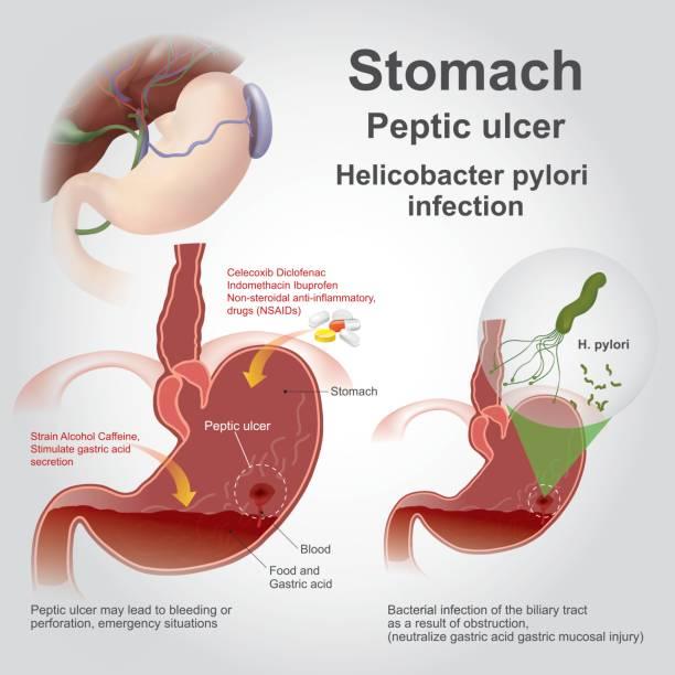
H. pylori infection often causes no symptoms. When symptoms do occur, they are usually related to gastritis or a peptic ulcer rather than cancer. Stomach cancer can also cause digestive symptoms, so symptoms alone cannot distinguish an H. pylori infection, an ulcer, or cancer.
When H. pylori causes symptoms, they may include:
Burning or gnawing stomach pain, sometimes worse when the stomach is empty
Bloating and indigestion
Nausea
Frequent belching
Reduced appetite
These symptoms are more commonly associated with gastritis or peptic ulcers than with stomach cancer. Many people with H. pylori infection have no symptoms at all.
Seek prompt medical evaluation if you experience:
Unexplained weight loss
Black, tarry stools or blood in the stool
Vomiting blood or coffee-ground-like vomit
Persistent vomiting or difficulty swallowing
Severe abdominal pain that does not improve with antacids
These symptoms can have causes other than cancer, but they should not be ignored. They warrant medical assessment and may require an upper endoscopy, rather than self-diagnosis. Endoscopy allows a doctor to examine the stomach lining and take biopsies when needed
Most peptic ulcers are benign and do not automatically become cancer. H. pylori infection and NSAID use are common causes of peptic ulcers. However, a gastric ulcer can sometimes be caused by an underlying cancer, so its appearance alone cannot reliably establish that it is benign.
The distinction is where the ulcer is located:
Gastric ulcers: Most are benign, but a small proportion are malignant or associated with underlying cancer. Published studies have reported malignancy rates ranging from approximately 2.4% to 21%, depending on the study population and clinical setting. A large cohort study published in Endoscopy International Open found that 6% of gastric ulcers were malignant, highlighting why gastric ulcers require appropriate biopsy and follow-up.
Duodenal ulcers: These are overwhelmingly benign. Cancer arising in a typical duodenal ulcer is extremely rare, so the management approach differs from that of a gastric ulcer.
Larger or “giant” gastric ulcers deserve particular attention. A 2018 retrospective cohort study by Lord et al., published in the United European Gastroenterology Journal, evaluated 111 patients with giant gastric ulcers measuring at least 3 cm. Forty-two ulcers were malignant, giving a malignancy yield of 37.8% (95% CI 28.8–46.8%). This was a selected high-risk population and should not be interpreted as the cancer risk for gastric ulcers in general. The findings nevertheless illustrate why ulcer size, location, appearance, and biopsy results matter.
A gastric ulcer that looks benign can occasionally conceal cancer. Endoscopy with biopsy is used to establish whether the ulcer is benign or malignant, rather than relying on appearance alone. Multiple samples from the ulcer edge and base may be taken, particularly when malignancy is suspected.
After treatment, repeat endoscopy is often recommended to confirm healing, especially when the ulcer appears suspicious, symptoms persist, or the initial biopsy is negative but concern remains. A negative biopsy does not completely exclude malignancy, and an ulcer that has not healed should be reassessed.
Bottom line: A stomach ulcer does not mean cancer, but it should be properly evaluated. A duodenal ulcer is generally benign, whereas a gastric ulcer requires more careful assessment.
A stomach ulcer and stomach cancer can cause similar digestive symptoms, but they are different conditions. Symptoms alone cannot reliably distinguish them. A gastric ulcer may be benign or may conceal an underlying malignancy, which is why appropriate evaluation matters.
|
Feature |
Stomach Ulcer |
Stomach Cancer |
|---|---|---|
|
What is it? |
An open sore in the stomach lining. |
Abnormal cells that grow uncontrollably in the stomach. |
|
Onset |
Pain may develop suddenly or gradually and may be related to meals or an empty stomach. |
Early stages may cause no symptoms. |
|
Pain pattern |
Burning or gnawing pain may vary with food timing and sometimes improves with antacids. |
Discomfort may be persistent or progressive and does not reliably follow the same pattern. |
|
Common symptoms |
Upper-abdominal pain, indigestion, nausea, bloating, or belching. |
Indigestion, upper-abdominal discomfort, nausea, or reduced appetite. |
|
Warning signs |
Bleeding can cause black stools or vomiting blood. |
Unexplained weight loss, persistent fatigue, and early satiety may occur. These are not unique to cancer. |
|
Main causes |
H. pylori infection and NSAID use are the most common causes. |
Multiple contributing factors, including H. pylori infection, smoking, diet, and family history. |
|
Can it become cancer? |
Most ulcers are benign. A gastric ulcer can sometimes conceal or be associated with cancer. |
Cancer is a malignant disease, not a benign ulcer. |
|
Diagnosis |
Endoscopy may be needed, especially for a gastric ulcer. Biopsy is performed when indicated. |
Endoscopy with biopsy establishes the diagnosis. CT and other tests help determine the stage. |
|
Treatment |
H. pylori eradication when present, acid-suppressing medication, and stopping the causative NSAID when appropriate. |
Oncology-led treatment may include surgery, chemotherapy, targeted therapy, immunotherapy, or radiation, depending on the cancer. |
|
Prognosis |
Most peptic ulcers heal with appropriate treatment. |
Prognosis depends on the cancer type, stage, and response to treatment. |
A benign gastric ulcer and a malignant ulcer can look similar on initial endoscopy. Endoscopy with biopsy is the reliable way to distinguish them, while imaging and other investigations help determine whether cancer is present and how far it has spread.
If you have persistent upper-abdominal symptoms, unexplained weight loss, black stools, vomiting blood, or difficulty swallowing, seek medical evaluation rather than assuming the cause is an ulcer.
H. pylori is a major cause of stomach cancer, but it is not the only risk factor. Diet, tobacco use, and family history can also influence gastric cancer risk. Having one or more risk factors does not mean that cancer will develop.
High intake of salt-preserved, pickled, or smoked foods is associated with increased stomach cancer risk and may contribute to stomach lining damage.
Smoking roughly doubles stomach cancer risk and may make H. pylori treatment less effective.
Having a first-degree relative with stomach cancer increases the risk of developing gastric cancer. The National Cancer Institute reports that seven studies found relative risks ranging from 1.5-fold to 3.5-fold among people with a first-degree relative affected by gastric cancer. These estimates come largely from retrospective studies and may be influenced by shared environmental factors, diet, and H. pylori exposure within families.
A Rapid Urease Test (RUT) is commonly performed during endoscopy to detect H. pylori in a stomach biopsy. Understanding what a positive result means can help avoid confusion between H. pylori infection and stomach cancer.
Helicobacter pylori can be diagnosed using a urea breath test, stool antigen test, blood antibody test, or endoscopy with biopsy. During endoscopy, a Rapid Urease Test (RUT) can be performed on a biopsy sample to detect active H. pylori infection.
No. A positive RUT result means H. pylori was detected in the biopsy sample. It confirms infection, not cancer.
RUT and histopathology are different tests that answer different questions. The pathologist examines the same or an adjacent biopsy sample under a microscope to look for cancerous or precancerous changes. A positive RUT does not establish malignancy, and a negative RUT does not rule it out.
What happens next? If H. pylori is confirmed, appropriate antibiotic eradication treatment can reduce future gastric cancer risk. Your doctor will also interpret any biopsy findings and decide whether further evaluation is needed.
Treating H. pylori infection can lower the future risk of stomach cancer, particularly when eradication occurs before advanced precancerous changes develop.
According to the American College of Gastroenterology, treatment typically combines multiple antibiotics with an acid-suppressing medicine such as a proton pump inhibitor (PPI). The exact regimen should be selected by a doctor based on factors such as previous antibiotic exposure and local resistance patterns.
Eradicating H. pylori can significantly reduce gastric cancer risk. Evidence suggests the benefit is greatest when treatment is given before extensive atrophic gastritis or intestinal metaplasia develops.
The American College of Gastroenterology recommends that treatment should be followed by a test of cure using a urea breath test, stool antigen test, or appropriate biopsy-based test at least 4 weeks after completing antibiotics. PPIs generally need to be stopped for about 2 weeks before testing to reduce the chance of a false-negative result, under medical guidance.
At MyDiagnostics, patients can access H. pylori testing options to help detect infection and monitor treatment response. Testing should be interpreted by a healthcare professional alongside symptoms and other clinical findings.
Get tested with MyDiagnostics and discuss your results with your healthcare provider for appropriate treatment and follow-up.
Medical Disclaimer: This content is for general information only and does not replace professional medical advice, diagnosis, or treatment. If you have persistent or concerning symptoms, consult a qualified healthcare professional.